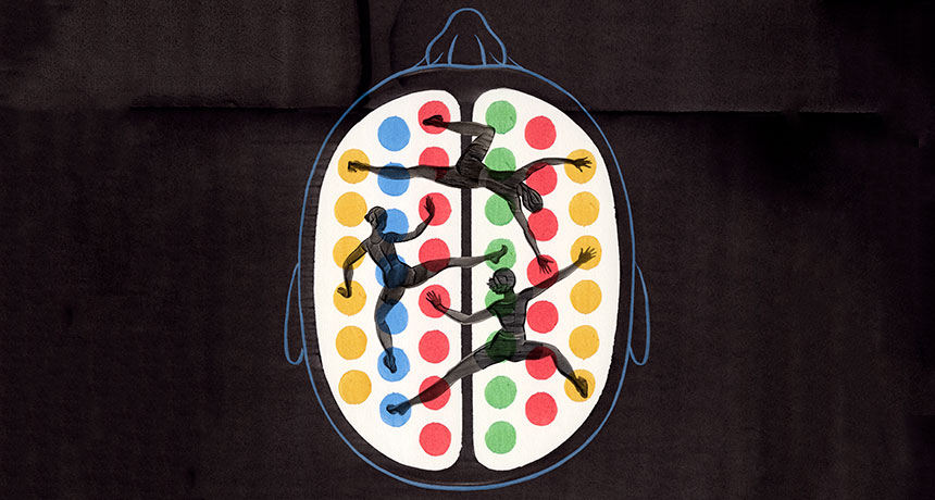
Learning takes brain acrobatics

Peer inside the brain of someone learning. You might be lucky enough to spy a synapse pop into existence. That physical bridge between two nerve cells seals new knowledge into the brain. As new information arrives, synapses form and strengthen, while others weaken, making way for new connections.

You might see more subtle changes, too, like fluctuations in the levels of signaling molecules, or even slight boosts in nerve cell activity. Over the last few decades, scientists have zoomed in on these microscopic changes that happen as the brain learns. And while that detailed scrutiny has revealed a lot about the synapses that wire our brains, it isn’t enough. Neuroscientists still lack a complete picture of how the brain learns.

They may have been looking too closely. When it comes to the neuroscience of learning, zeroing in on synapse action misses the forest for the trees.

A new, zoomed-out approach attempts to make sense of the large-scale changes that enable learning. By studying the shifting interactions between many different brain regions over time, scientists are beginning to grasp how the brain takes in new information and holds onto it.
These kinds of studies rely on powerful math. Brain scientists are co-opting approaches developed in other network-based sciences, borrowing tools that reveal in precise, numerical terms the shape and function of the neural pathways that shift as human brains learn.

“When you’re learning, it doesn’t just require a change in activity in a single region,” says Danielle Bassett, a network neuroscientist at the University of Pennsylvania. “It really requires many different regions to be involved.” Her holistic approach asks, “what’s actually happening in your brain while you’re learning?” Bassett is charging ahead to both define this new field of “network neuroscience” and push its boundaries.

“This line of work is very promising,” says neuroscientist Olaf Sporns of Indiana University Bloomington. Bassett’s research, he says, has great potential to bridge gaps between brain-imaging studies and scientists’ understanding of how learning happens. “I think she’s very much on the right track.”
Already, Bassett and others have found tantalizing hints that the brains that learn best have networks that are flexible, able to rejigger connections on the fly to allow new knowledge in. Some brain regions always communicate with the same neural partners, rarely switching to others. But brain regions that exhibit the most flexibility quickly swap who they’re talking with, like a parent who sends a birthday party invite to the preschool e-mail list, then moments later, shoots off a work memo to colleagues.

In a few studies, researchers have witnessed this flexibility in action, watching networks reconfigure as people learn something while inside a brain scanner. Network flexibility may help several types of learning, though too much flexibility may be linked to disorders such as schizophrenia, studies suggest.

Not surprisingly, some researchers are rushing to apply this new information, testing ways to boost brain flexibility for those of us who may be too rigid in our neural connections.

“These are pretty new ideas,” says cognitive neuroscientist Raphael Gerraty of Columbia University. The mathematical and computational tools required for this type of research didn’t exist until recently, he says. So people just weren’t thinking about learning from a large-scale network perspective. “In some ways, it was a pretty boring mathematical, computational roadblock,” Gerraty says. But now the road is clear, opening “this conceptual avenue … that people can now explore.”
It takes a neural village
That conceptual avenue is more of a map, made of countless neural roads. Even when a person learns something very simple, large swaths of the brain jump in to help. Learning an easy sequence of movements, like tapping out a brief tune on a keyboard, prompts activity in the part of the brain that directs finger movements. The action also calls in brain areas involved in vision, decision making, memory and planning. And finger taps are a pretty basic type of learning. In many situations, learning calls up even more brain areas, integrating information from multiple sources, Gerraty says.

He and colleagues caught glimpses of some of these interactions by scanning the brains of people who had learned associations between two faces. Only one of the faces was then paired with a reward. In later experiments, the researchers tested whether people could figure out that the halo of good fortune associated with the one face also extended to the face it had been partnered with earlier. This process, called “transfer of learning,” is something that people do all the time in daily life, such as when you’re wary of the salad at a restaurant that recently served tainted cheese.

Study participants who were good at applying knowledge about one thing — in this case, a face — to a separate thing showed particular brain signatures, Gerraty and colleagues reported in 2014 in the Journal of Neuroscience. Connections between the hippocampus, a brain structure important for memory, and the ventromedial prefrontal cortex, involved in self-control and decision making, were weaker in good learners than in people who struggled to learn. The scans, performed several days after the learning task, revealed inherent differences between brains, the researchers say. The experiment also turned up other neural network differences among these regions and larger-scale networks that span the brain.

Children who have difficulty learning math, when scanned, also show unexpected brain connectivity, according to research by neuroscientist Vinod Menon of Stanford University and colleagues. Compared with kids without disabilities, children with developmental dyscalculia who were scanned while doing math problems had more connections, particularly among regions involved in solving math problems. That overconnectivity, described in 2015 in Developmental Science, was a surprise, Menon says, since earlier work had suggested that these math-related networks were too weak. But it may be that too many links create a system that can’t accommodate new information. “The idea is that if you have a hyperconnected system, it’s not going to be as responsive,” he says.
There’s a balance to be struck, Menon says. Neural pathways that are too weak can’t carry necessary information, and pathways that are too connected won’t allow new information to move in. But the problem isn’t as simple as that. “It’s not that everything is changing everywhere,” he says. “There is a specificity to it.” Some connections are more important than others, depending on the task.

Neural networks need to shuttle information around quickly and fluidly. To really get a sense of this movement as opposed to snapshots frozen in time, scientists need to watch the brain as it learns. “The next stage is to figure out how the networks actually shift,” Menon says. “That’s where the studies from Dani Bassett and others will be very useful.”

Flexing in real time
Bassett and colleagues have captured these changing networks as people learn. Volunteers were given simple sequences to tap out on a keyboard while undergoing a functional MRI scan. During six weeks of scanning as people learned the task, neural networks in their brains shifted around. Some connections grew stronger and some grew weaker, Bassett and her team reported in Nature Neuroscience in 2015.

People who quickly learned to tap the correct sequence of keys showed an interesting neural trait: As they learned, they shed certain connections between their frontal cortex, the outermost layer of the brain toward the front of the head, and the cingulate, which sits toward the middle of the brain. This connection has been implicated in directing attention, setting goals and making plans, skills that may be important for the early stages of learning but not for later stages, Bassett and colleagues suspect. Compared with slow learners, fast learners were more likely to have shunted these connections, a process that may have made their brains more efficient.

Flexibility seems to be important for other kinds of learning too. Reinforcement learning, in which right answers get a thumbs up and wrong answers are called out, also taps into brain flexibility, Gerraty, Bassett and others reported online May 30 at bioRxiv.org. This network comprises many points on the cortex, the brain’s outer layer, and a deeper structure known as the striatum. Other work on language comprehension, published by Bassett and colleagues last year in Cerebral Cortex, found some brain regions that were able to quickly form and break connections.

These studies captured brains in the process of learning, revealing “a much more interesting network structure than what we previously thought when we were only looking at static snapshots,” Gerraty says. The learning brain is incredibly dynamic, he says, with modules breaking off from partners and finding new ones.

While the details of those dynamics differ from study to study, there is an underlying commonality: “It seems that part of learning about the world is having parts of your brain become more flexible, and more able to communicate with different areas,” Gerraty says. In other words, the act of learning takes flexibility.

But too much of a good thing may be bad. While performing a recall task in a scanner, people with schizophrenia had higher flexibility among neural networks across the brain than did healthy people, Bassett and colleagues reported last year in the Proceedings of the National Academy of Sciences. “That suggests to me that while flexibility is good for healthy people, there is perhaps such a thing as too much flexibility,” Bassett says.
Just how this flexibility arises, and what controls it, is unknown. Andrea Stocco, a cognitive neuroscientist at the University of Washington in Seattle, suspects that a group of brain structures called the basal ganglia, deep within the brain, has an important role in controlling flexibility. He compares this region, which includes the striatum, to an air traffic controller who shunts information to where it’s most needed. One of the basal ganglia’s jobs seems to be shutting things down. “Most of the time, the basal ganglia is blocking something,” he says. Other researchers have found evidence that crucial “hubs” in the cortex help control flexibility.

Push for more
Researchers don’t yet know how measures of flexibility in brain regions relate to the microscopic changes that accompany learning. For now, the macro and the micro views of learning are separate worlds. Despite that missing middle ground, researchers are charging ahead, looking for signs that neural flexibility might offer a way to boost learning aptitude.

It’s possible that external brain stimulation may enhance flexibility. After receiving brain stimulation carefully aimed at a known memory circuit, people were better able to recall lists of words, scientists reported May 8 in Current Biology. If stimulation can boost memory, some argue, the technique could enhance flexibility and perhaps learning too.
Certain drugs show promise. DXM, found in some cough medicines, blocks proteins that help regulate nerve cell chatter. Compared with a placebo, the compound made some brain regions more flexible and able to rapidly switch partners in healthy people, Bassett and colleagues reported last year in the Proceedings of the National Academy of Sciences. She is also studying whether neurofeedback — a process in which people try to change their brain patterns to become more flexible with real-time monitoring — can help.

Something even simpler might work for boosting flexibility. On March 31 in Scientific Reports, Bassett and colleagues described their network analyses of an unusual subject. For a project called MyConnectome, neuroscientist Russ Poldrack, then at the University of Texas at Austin, had three brain scans a week for a year while assiduously tracking measures that included mood. Bassett and her team applied their mathematical tools to Poldrack’s data to get measurements of his neural flexibility on any given scan day. The team then looked for associations with mood. The standout result: When Poldrack was happiest, his brain was most flexible, for reasons that aren’t yet clear. (Flexibility was lowest when he was surprised.)

Those results are from a single person, so it’s unknown how well they would generalize to others. What’s more, the study identifies only a link, not that happiness causes more flexibility or vice versa. But the idea is intriguing, if not obvious, Bassett says. “Of course, no teacher is really going to say we’re doing rocket science if we tell them we should make the kids happier and then they’ll learn better.” But finding out exactly how happiness relates to learning is important, she says.

The research is just getting started. But already, insights on learning are coming quickly from the small group of researchers viewing the brain as a matrix of nodes and links that deftly shift, swap and rearrange themselves. Zoomed out, network science brings to the brain “a whole new set of hypotheses and new ways of testing them,” Bassett says.

Microbes hobble a widely used chemo drug

Some bacteria may shield tumor cells against a common chemotherapy drug.

Certain types of bacteria make an enzyme that inactivates the drug gemcitabine, researchers report in the Sept. 15 Science. Gemcitabine is used to treat patients with pancreatic, lung, breast and bladder cancers.

Bacteria that produce the enzyme cytidine deaminase converted the drug to an inactive form. That allowed tumor cells to survive gemcitabine treatment in lab dishes and mouse studies, Leore Geller of the Weizmann Institute of Science in Rehovot, Israel, and colleagues discovered. More than 98 percent of the enzyme-producing microbes belong to the Gammaproteobacteria class, which includes E. coli and about 250 bacterial genera.
Pancreatic tumors taken from human patients also carried the enzyme-producing bacteria. Of 113 pancreatic ductal adenocarcinoma samples studied, 86 contained gemcitabine-inactivating bacteria.

Antibiotics may correct the problem. In the study, Geller and colleagues infected mice that had colon cancer with the enzyme-producing bacteria. Tumors grew rapidly in infected mice treated with gemcitabine alone. Giving the mice antibiotics helped gemcitabine kill tumor cells, increasing the number of tumor cells going through a type of cell death called apoptosis from about 15 percent to 60 percent or more. That result may indicate that combinations of gemcitabine and antibiotics could make chemotherapy more effective for some cancer patients.

Body clock mechanics wins U.S. trio the Nobel Prize in physiology or medicine

Discoveries about the clocklike ups and downs of daily life have won Jeffery C. Hall, Michael Rosbash and Michael W. Young the Nobel Prize in physiology or medicine.

Circadian rhythms are daily cycles of hormones, gene activity and other biological processes that govern sleep, body temperature and metabolism. When thrown out of whack, there can be serious health consequences, including increased risk of diabetes, heart and Alzheimer’s diseases.

Hall and Rosbash discovered the first molecular gear of the circadian clockworks: A protein called Period increases and decreases in abundance on a regular cycle during the day. Young discovered that another protein called Timeless works with Period to drive the clock. Young also discovered other circadian clockworks.

Nanoscale glitches let flowers make a blue blur that bees can see

A bit of imperfection could be perfect for flowers creating a “blue halo” effect that bees can see.

At least a dozen families of flowering plants, from hibiscuses to daisy relatives, have a species or more that can create a bluish-ultraviolet tinge using arrays of nanoscale ridges on petals, an international research team reports online October 18 in Nature. These arrays could be the first shown to benefit from the sloppiness of natural fabrication, says coauthor Silvia Vignolini, a physicist specializing in nanoscale optics at the University of Cambridge.
Flowers, of course, can’t reach industrial standards for uniform nanoscale fabrication. Yet the halo may be a case where natural imperfections may be important to a flower’s display. Tests with artificial flowers showed that the nanoglitches made it easier for bees to learn that a showy petal meant a sugary reward, Vignolini and colleagues found.
Blues are rare in actual pigments in living things( SN: 12/10/16, p. 4 ). Color in the wings of Morpho butterflies or blue jay feathers, for instance, comes from nanoscale structures that contain no pigments but create colorful illusions by muting some wavelengths of light while intensely reflecting others ( SN: 6/11/16, p. 32 ).
Flower petals make their blue halo illusion with somewhat irregular versions of what are called diffraction gratings, rows of ridges like the recording surface on a CD. A perfectly regular array of ridges would create true iridescence, changing color depending on the angle a viewer takes. The flowers’ imperfections, variations in ridge height and spacing, weaken or destroy the iridescence. A viewer swooping by would see less color shifting and more of a bluish-ultraviolet tinge reflected at a wider range of angles.

To see whether bees respond more to iridescence or a blue halo, researchers created sets of artificial flowers, pieces of epoxy resin with some kind of nanoscale-ridged array. A petal-scale project was huge compared with the usual nanoscale experiments, requiring marathon fabrication sessions. “We were a pain to everybody,” Vignolini says.

In two tests, researchers offered bumblebees a pair of “flowers,” one that held sugar water and one with a nasty-tasting solution, to see how quickly bees would learn to distinguish sweet from foul. When the flower’s nanoridges had imperfections creating a blue halo, bees learned the task faster than when the flower had perfect iridescence. Imperfect arrays were actually an advantage for the flowers in creating displays pollinating bees find memorable, the researchers conclude.
Such disorder in nature’s structural color (versus pigments) has shown up before, as in obviously jumbled color-trick structures in bird feathers. Before the tests, though, it was unclear whether flowers would benefit from perfect iridescence and were just falling short in growing perfect arrays. The blue halo might have been merely a side effect of challenging botanical fabrication. The bee experiments, however, showed the opposite, the researchers say. These are the first tests to show that some disorder is not just a downside of natural fabrication but in itself “has a function,” Vignolini says.

That result makes sense to visual ecologist Nathan Morehouse of the University of Cincinnati. Nanostructures that iridesce may often just be a way birds or butterflies can create an unusual color rather than a way to produce iridescence for its own sake. The shifting colors might even have a downside. By definition, true iridescence changes color as an insect or bird changes its angle of approach, and so may not be the best form for an easy-to-remember signal. “Iridescence itself is something they just have to manage,” he suggests.

Alligators eat sharks — and a whole lot more

Alligators don’t just stick to freshwater and the prey they find there. These crafty reptiles can live quite easily, at least for a bit, in salty waters and find plenty to eat — including crabs, sea turtles and even sharks.

“They should change the textbooks,” says James Nifong, an ecologist with the Kansas Cooperative Fish and Wildlife Research Unit at Kansas State University in Manhattan, who has spent years documenting the estuarine gator diet.

Nifong’s most recent discovery, splashed all over the news last month, is that the American alligator (Alligator mississippiensis) eats at least three species of shark and two species of rays, he and wildlife biologist Russell Lowers report in the September Southeastern Naturalist.

Lowers captured a female gator with a young Atlantic stingray in her jaws near where he works at Kennedy Space Center in Cape Canaveral, Florida. And he and Nifong gathered several other eyewitness accounts: A U.S. Fish and Wildlife employee spotted a gator consuming a nurse shark in a Florida mangrove swamp in 2003. A birder photographed an alligator eating a bonnethead shark in a Florida salt marsh in 2006. One of Nifong’s collaborators, a marine turtle researcher, saw gators consuming both bonnethead and lemon sharks in the late 1990s. And Nifong found yet another report of a gator eating a bonnethead shark in Hilton Head, S.C., after their paper was published. All of these snacks required gators to venture into salty waters.
But shark may not be the most surprising item on the alligator estuarine menu. Nifong spent years catching hundreds of wild gators and pumping their stomachs to figure out what they eat, work that relies “on electrical tape, duct tape and zip ties,” Nifong says. And he found that the menu is pretty long.

To snag an alligator, he uses a big blunted hook or, with smaller animals, just grabs the animal and hauls it into the boat. He gets a noose around its neck. Then the researchers tape the mouth shut, take body measurements (everything from weight to toe length) and get blood or urine samples.

Once that’s out of the way, the team will strap the gator to a board with Velcro ties or rope. Then, it’s time to untape the mouth, quickly insert a piece of pipe to hold it open, and tape the alligator’s mouth around the pipe. The pipe, Nifong says, is there “so they can’t bite down.” And that’s important, because next someone has to stick a tube down the gator’s throat and hold it there to keep the animal’s throat open.
Finally, “we fill [the stomach] up with water very slowly so we don’t injure the animal,” Nifong says. “Then we do basically the Heimlich maneuver.” Pressing down on the abdomen forces the gator to give up its stomach contents. Usually.
“Sometimes it goes better than other times,” he says. “They can just decide to not let it out.” Then the researchers carefully undo all their work to let the gator loose.

Back in the lab, Nifong and his colleagues teased out what they could find in those stomach contents, and looked for more clues about the animals’ diet from in the blood samples. Nifong and his colleagues found that the gators were eating a rich marine diet, including small fish, mammals, birds, insects and crustaceans. They’ll even eat fruit and seeds. The sharks and rays didn’t show up in these studies (nor did sea turtles, which gators have also been spotted munching on). But Nifong and Lowers speculate that’s because the tissue of those animals gets digested very quickly. So if a gator had eaten a shark more than a few days before being caught, there was no way to know.

Because alligators don’t have any salt glands, “they’re subject to the same pressures as me or you when being out in saltwater,” Nifong says. “You’re losing water, and you’re increasing salt in your blood system.” That can lead to stress and even death, he notes. So the gators tend to just go back and forth between saltwater and freshwater. They can also close off their throat with a cartilaginous shield and shut their nostrils to keep salty water out. And when they eat, they’ll tip their head up to let the saltwater drain out before gulping down their catch.
What alligators eat isn’t as important a finding as the discovery that they regularly travel between saltwater and freshwater environments, Nifong says. And, he notes, “it occurs across a wide variety of habitats across the U.S. southeast.” That’s important because the gators are moving nutrients from rich marine waters into poorer, fresh waters. And they may be having a larger effect on estuarine food webs that anyone had imagined.

For instance, one of the prey items on the alligator menu is blue crab. Gators “scare the bejesus out of them,” Nifong says. And when gators are around, blue crabs decrease their predation of snails, which might then eat more of the cordgrass that forms the base of the local ecosystem. “Understanding that an alligator has a role in that kind of interaction,” Nifong points out, is important when planning conservation efforts.

Artificial insulin-releasing cells may make it easier to manage diabetes

Artificial cells made from scratch in the lab could one day offer a more effective, patient-friendly diabetes treatment.

Diabetes, which affects more than 400 million people around the world, is characterized by the loss or dysfunction of insulin-making beta cells in the pancreas. For the first time researchers have created synthetic cells that mimic how natural beta cells sense blood sugar concentration and secrete just the right amount of insulin. Experiments with mice show that these cells can regulate blood sugar for up to five days, researchers report online October 30 in Nature Chemical Biology.
If the mouse results translate to humans, diabetics could inject these artificial beta cells to automatically regulate their blood sugar levels for days at a time.

That would be a “a huge leap forward” for diabetic patients who currently have to check their blood sugar and inject insulin several times a day, says Omid Veiseh, a bioengineer at Rice University in Houston who wasn’t involved in the research. “Even if it were just a one-day thing, it would still be impressive,” he says.
Fashioned from human-made materials and biological ingredients like proteins, these faux cells contain insulin-filled pouches much like the insulin-carrying compartments inside real beta cells. And, similar to a natural beta cell, when one of these artificial beta cells is surrounded by excess blood sugar, its insulin sacs fuse with its outer membrane and eject insulin into the bloodstream. As blood sugar levels drop, insulin packets stop fusing with the membrane, which stems the cell’s insulin secretion.
Fabricating artificial insulin delivery systems that actually imitate the inner workings of real beta cells for ultrafine blood sugar regulation is “an engineering feat,” says Patrik Rorsman, a diabetes researcher at the University of Oxford who wasn’t involved in the work. The cellular imitations are “not as perfect as the beta cells we’re equipped with when we’re healthy,” he adds. For one thing, the faux cells eventually run out of insulin to release. But Rorsman believes that such artificial cells present a viable diabetes treatment.
Unlike transplanted beta cells — or other types of real cells genetically engineered to release insulin for diabetes treatment (SN: 1/15/11, p. 9) — these artificial cells could be mass-produced and have a much longer shelf life than live cells, says study coauthor Zhen Gu, a biomedical engineer at the University of North Carolina at Chapel Hill.

When Gu and colleagues injected their synthetic cells into diabetic mice, the animals’ blood sugar levels normalized within an hour and stayed that way up to five days, when the cells ran out of insulin. The researchers plan to perform further tests on lab animals to assess the fake cells’ long-term health effects before running clinical trials.

Even for patients who manage their insulin with automated mechanical pumps (SN Online: 5/8/10), synthetic cells offer the advantage of more precise, real time blood sugar regulation, says Michael Strano, a bioengineer at MIT. The creation of the new faux cells not only poses a potential diabetes treatment, “but it’s also a bellwether. It’s slightly ahead of its time,” says Strano. “I think therapeutics of the future are going to look like this.”

Studying giant tortoise flips without tipping the animals over is a delicate business

It would be a memorable sight. But it would also be so wrong to tip over Galápagos giant tortoises to see how shell shape affects their efforts to leg-pump, neck-stretch and rock right-side up again.

Shell shape matters, says evolutionary biologist Ylenia Chiari, though not the way she expected. It’s taken years, plus special insights from a coauthor who more typically studies scorpions, for Chiari and her team to measure and calculate their way to that conclusion. But no endangered species have been upended in the making of the study.
“They’re amazing,” says Chiari of the dozen or so species of Chelonoidis grazing over the Galápagos Islands. Hatchlings start not quite the size of a tennis ball and after decades, depending on species and sex, “could be like — a desk,” says Chiari, of the University of South Alabama in Mobile.

Two extremes among the species’ shell shapes intrigue Chiari: high-domed mountains versus mere hillocks called saddlebacks because of an upward flare saddling the neck. Researchers have dreamed up possible benefits for the shell differences, such as the saddleback flare letting tortoises stretch their necks higher upward in grazing on sparse plants.
At the dryer, lower altitudes where saddleback species tend to live, fields of lava chunks and cacti make walking treacherous. “I fell on a cactus once,” Chiari says. Tortoises tumble over, too, and she wondered whether saddleback shells might be easier to set right again.
She went paparazzi on 89 tortoise shells, taking images from multiple angles to create a 3-D computerized version of each shell. Many shells were century-old museum specimens from the California Academy of Sciences in San Francisco, but she stalked some in the wild, too. The domed tortoises tended to pull into their shells with a huffing noise during their time in front of the lens and just wait till the weirdness ended. A saddleback species plodded toward the interruption, though, butting and biting (toothless but emphatic) at her legs.

To calculate energy needed to rock and roll the two shell types, Chiari needed to know the animals’ centers of mass. No one, however, had measured it for any tortoise. Enter coauthor Arie van der Meijden of CIBIO, Research Center in Biodiversity and Genetic Resources at the University of Porto in Portugal. With expertise in biomechanics, he scaled up from the arthropods he often studies. For a novel test of tortoises, he arranged for a manufacturer to provide equipment measuring force exerted at three points under a tiltable platform. As the first giant tortoise, weighing in at about 100 kilograms, started to lumber aboard the platform at Rotterdam’s zoo, Chiari thought, “Oh my gosh, it’s going to crush everything.” For a gentler and more even landing, four men heaved the tortoise into position.

Calculating the centers of mass for Rotterdam tortoises, the researchers extrapolated to the 89 shells. The low, flattened saddleback shape actually made shells tougher to right, taking more energy, the team reports November 30 in Scientific Reports. Now Chiari muses over whether the saddle at the shell front might let freer neck movements compensate after a trip and a flip.

Jackpot of fossilized pterosaur eggs unearthed in China

Hundreds of eggs belonging to a species of flying reptile that lived alongside dinosaurs are giving scientists a peek into the earliest development of the animals.

The find includes at least 16 partial embryos, several still preserved in 3-D. Those embryos suggest that the animals were able to walk, but not fly, soon after hatching, researchers report in the Dec. 1 Science.

Led by vertebrate paleontologist Xiaolin Wang of the Chinese Academy of Sciences in Beijing, the scientists uncovered at least 215 eggs in a block of sandstone about 3 meters square. All of the eggs belonged to one species of pterosaur, Hamipterus tianshanensis, which lived in the early Cretaceous Period about 120 million years ago in what is now northwestern China.
Previously, researchers have found only a handful of eggs belonging to the winged reptiles, including five eggs from the same site in China (SN: 7/12/14, p. 20) and two more found in Argentina. One of the Argentinian eggs also contained a flattened but well-preserved embryo.
One reason for the dearth of fossils may be that the eggs were rather soft with a thin outer shell, unlike the hard casings of eggs belonging to dinosaurs, birds and crocodiles but similar to those of modern-day lizards. Due to that soft shape, pterosaur eggs also tend to flatten during preservation. Finding fossilized eggs containing 3-D embryos opens a new window into pterosaur development, says coauthor Alexander Kellner, a vertebrate paleontologist at Museu Nacional/Universidade Federal do Rio de Janeiro.
The eggs weren’t found at an original nesting site but had been jumbled and deformed, probably transported by a flood during an intense storm, Kellner says. Sand and other sediments carried by the water would then have rapidly buried the soft eggs, which was necessary to preserve them, Kellner says. “Otherwise, they would have decomposed.”
Using computerized tomography, the researchers scanned the internal contents of the eggs. Two of the best-preserved embryos revealed a tantalizing clue to pterosaur development, Kellner says. A key part of a wing bone, called the deltopectoral crest, was not fully developed in the embryos, even in an embryo the researchers interpret as nearly at term. The femur, or leg bone, of the embryo, however, was well developed. This suggests that, when born, the hatchlings could walk but not yet fly and may have still required some parental care for feeding, the scientists propose.
Such an interpretation requires an abundance of caution, says D. Charles Deeming, a vertebrate paleontologist at the University of Lincoln in England not involved in the study. For example, he says, there isn’t enough evidence to say for certain that the embryo in question was nearly at term and, therefore, to say that it couldn’t fly when born, a point he also raises in a column published in the same issue of Science. “There’s a real danger of overinterpretation.” But with such a large group of eggs, he says, researchers can make quantitative measurements to better understand the range of egg sizes and shapes to get a sense of variation in animal size.

Kellner says this work is ongoing and agrees that there is still a significant amount of study to be done on these and other eggs more recently found at the site. And the hunt is on for more concentrations of eggs in the same site. “Now that we know what they look like, we can go back and find more. You just have to get your knees down and look.”

AI eavesdrops on dolphins and discovers six unknown click types

A new computer program has an ear for dolphin chatter.

The algorithm uncovered six previously unknown types of dolphin echolocation clicks in underwater recordings from the Gulf of Mexico, researchers report online December 7 in PLOS Computational Biology. Identifying which species produce the newly discovered click varieties could help scientists better keep tabs on wild dolphin populations and movements.

Dolphin tracking is traditionally done with boats or planes, but that’s expensive, says study coauthor Kaitlin Frasier, an oceanographer at the Scripps Institution of Oceanography in La Jolla, Calif. A cheaper alternative is to sift through seafloor recordings — which pick up the echolocation clicks that dolphins make to navigate, find food and socialize. By comparing different click types to recordings at the surface — where researchers can see which animals are making the noise — scientists can learn what different species sound like, and use those clicks to map the animals’ movements deep underwater.
But even experts have trouble sorting recorded clicks, because the distinguishing features of these signals are so subtle. “When you have analysts manually going through a dataset, then there’s a lot of bias introduced just from the human perception,” says Simone Baumann-Pickering, a biologist at the Scripps Institution of Oceanography not involved in the work. “Person A may see things differently than person B.” So far, scientists have only determined the distinct sounds of a few species.
To sort clicks faster and more precisely, Frasier and her colleagues outsourced the job to a computer. They fed an algorithm 52 million clicks recorded over two years by near-seafloor sound sensors across the Gulf of Mexico. The algorithm grouped echolocation clicks based on similarities in speed and pitch — the same criteria human experts use to classify clicks. “We don’t tell it how many click types to find,” Frasier says. “We just kind of say, ‘What’s in here?’”
The algorithm picked out seven major kinds of clicks, which the researchers think are made by different dolphin species. Frasier’s team recognized one class as being made by a species called Risso’s dolphin. The scientists suspect that another group of clicks, most common in recordings near the Green Canyon south of Louisiana, was produced by short-finned pilot whales that frequent this region. Another type resembles sounds from the eastern Pacific Ocean that a dolphin called the false killer whale makes.
To confirm the identifications, the researchers now need to compare their computer-generated categories against surface observations of these dolphins, Frasier says.

The algorithm’s click classes may not match up with dolphin species one-to-one, says Baumann-Pickering. If that were the case, “we would expect to see a heck of a lot more categories, really, based on the number of species that ought to be in that area,” she says. That absence suggests that some closely related species produce highly similar clicks the algorithm didn’t tease apart.

Still, “it would be great to be able to confidently assign certain species to each of the different click types, even if more than one species is assigned to a single click type,” says Lynne Hodge, a marine biologist at Duke University who wasn’t involved in the work. More precisely monitoring dolphins with seafloor recordings could provide new insight into how these animals respond to environmental problems such as oil spills and the long-term effects of climate change.

A quantum communications satellite proved its potential in 2017

During the world’s first telephone call in 1876, Alexander Graham Bell summoned his assistant from the other room, stating simply, “Mr. Watson, come here. I want to see you.” In 2017, scientists testing another newfangled type of communication were a bit more eloquent. “It is such a privilege and thrill to witness this historical moment with you all,” said Chunli Bai, president of the Chinese Academy of Sciences in Beijing, during the first intercontinental quantum-secured video call.

The more recent call, between researchers in Austria and China, capped a series of milestones reported in 2017 and made possible by the first quantum communications satellite, Micius, named after an ancient Chinese philosopher (SN: 10/28/17, p. 14).
Created by Chinese researchers and launched in 2016, the satellite is fueling scientists’ dreams of a future safe from hacking of sensitive communiqués. One day, impenetrable quantum cryptography could protect correspondences. A secret string of numbers known as a quantum key could encrypt a credit card number sent over the internet, or encode the data transmitted in a video call, for example. That quantum key would be derived by measuring the properties of quantum particles beamed down from such a satellite. Quantum math proves that any snoops trying to intercept the key would give themselves away.

“Quantum cryptography is a fundamentally new way to give us unconditional security ensured by the laws of quantum physics,” says Chao-Yang Lu, a physicist at the University of Science and Technology of China in Hefei, and a member of the team that developed the satellite.

But until this year, there’s been a sticking point in the technology’s development: Long-distance communication is extremely challenging, Lu says. That’s because quantum particles are delicate beings, easily jostled out of their fragile quantum states. In a typical quantum cryptography scheme, particles of light called photons are sent through the air, where the particles may be absorbed or their properties muddled. The longer the journey, the fewer photons make it through intact, eventually preventing accurate transmissions of quantum keys. So quantum cryptography was possible only across short distances, between nearby cities but not far-flung ones.

With Micius, however, scientists smashed that distance barrier. Long-distance quantum communication became possible because traveling through space, with no atmosphere to stand in the way, is much easier on particles.
In the spacecraft’s first record-breaking accomplishment, reported June 16 in Science, the satellite used onboard lasers to beam down pairs of entangled particles, which have eerily linked properties, to two cities in China, where the particles were captured by telescopes (SN: 8/5/17, p. 14). The quantum link remained intact over a separation of 1,200 kilometers between the two cities — about 10 times farther than ever before. The feat revealed that the strange laws of quantum mechanics, despite their small-scale foundations, still apply over incredibly large distances.

Next, scientists tackled quantum teleportation, a process that transmits the properties of one particle to another particle (SN Online: 7/7/17). Micius teleported photons’ quantum properties 1,400 kilometers from the ground to space — farther than ever before, scientists reported September 7 in Nature. Despite its sci-fi name, teleportation won’t be able to beam Captain Kirk up to the Enterprise. Instead, it might be useful for linking up future quantum computers, making the machines more powerful.

The final piece in Micius’ triumvirate of tricks is quantum key distribution — the technology that made the quantum-encrypted video chat possible. Scientists sent strings of photons from space down to Earth, using a method designed to reveal eavesdroppers, the team reported in the same issue of Nature. By performing this process with a ground station near Vienna, and again with one near Beijing, scientists were able to create keys to secure their quantum teleconference. In a paper published in the Nov. 17 Physical Review Letters, the researchers performed another type of quantum key distribution, using entangled particles to exchange keys between the ground and the satellite.

The satellite is “a major development,” says quantum physicist Thomas Jennewein of the University of Waterloo in Canada, who is not involved with Micius. Although quantum communication was already feasible in carefully controlled laboratory environments, the Chinese researchers had to upgrade the technology to function in space. Sensitive instruments were designed to survive fluctuating temperatures and vibrations on the satellite. Meanwhile, the scientists had to scale down their apparatus so it would fit on a satellite. “This has been a grand technical challenge,” Jennewein says.

Eventually, the Chinese team is planning to launch about 10 additional satellites, which would fly in formation to allow for coverage across more areas of the globe.